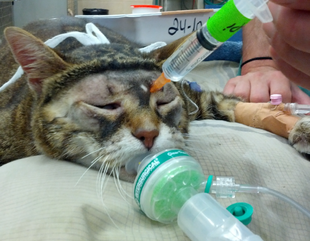
In this post, Yael Shilo-Benjamini DVM, DACVAA discusses the technique of providing peribulbar anesthesia, an alternative to retrobulbar anesthesia that may be safer and more effective.
Dr. Shilo-Benjamini completed an anesthesia and pain management residency at the University of California Davis, and is a clinical lecturer of anesthesia and analgesia at Koret School of Veterinary Medicine, The Hebrew University of Jerusalem. She has a particular interest in perioperative analgesia, especially with the use of regional anesthetic techniques.

Yael Shilo-Benjamini, DVM, Dip. ACVAA
Peribulbar anesthesia (PBA) is a simple, inexpensive, and relatively safe and effective regional anesthesia technique. In human ophthalmic surgery, it has replaced the use of retrobulbar anesthesia because of the advantage of limited risk of injury to intraconal structures, such as the optic nerve and major blood vessels. The primary indication to use PBA is enucleation, but in people it is also used for cataract and other intraocular surgeries.
PBA is performed by introducing a needle outside of the extraocular muscle cone (extraconal injection; Figure 1). I tend to use 25-gauge, 5/8-inch (1.6 cm) in cats and small-breed dogs, and 23-gauge, 1-inch (2.5 cm) in medium to large-breed dogs. In giant-breed dogs a longer needle, such as 22-23-gauge, 1.5-inch (3.8 cm), might be required.

In cats, 3.5-4 mL should be used per adult cat, but in kittens, I use approximately 1 mL per kg (up to 4 mL).
In dogs, a volume of 0.2-0.8 mL/kg was reported. It is important to note that the lower dosage should be used in larger-breed dogs, and the higher dosage in tiny breeds, due to changes in body surface area to volume ratio.
Prior to enucleation surgeries, it is advisable to use longer-acting local anesthetics, such as bupivacaine or ropivacaine. Many times (especially in cats and small-breed dogs) these drugs should be diluted with saline 0.9% to avoid toxicity. Calculate the maximum recommended dose (2 mg/kg for bupivacaine and 3 mg/kg for ropivacaine) then to dilute the local anesthetic to the required volume.
The insertion point of the needle in cats is at the dorso-medial orbit (Figure 2), while in dogs several insertions were reported: ventro-lateral orbit, dorso-medial + ventro-lateral orbit (Figure 3), or medial cantus (Figure 4). In all cases, the needle is inserted in close proximity to the orbital wall (I tend to scratch the wall), in order to avoid damaging any vessels and nerves, or penetrating into the globe.


Injection is performed after assuring a negative pressure with the syringe plunger, and while the bevel of the needle is facing the globe. In addition, because the large volume required, the rate of injection should be slow and a slight pressure should be applied to the needle in order to make sure that it will not move backward during injection.
Following injection, gentle massage of the eyelids can promote local anesthetic distribution.
Commonly reported complications of PBA include exophthalmos, chemosis (conjunctival edema), and ecchymosis (conjunctival bleeding due to spread of the local anesthetic into the eyelids and damage to minor blood vessels). These complications usually resolve within several hours after injection. In cats, a clinical important increase in IOP was reported following PBA, however, IOP decreased within 10 minutes from injection. When enucleation is planned, the increase in IOP is not a concern, but PBA should probably be avoided in animals with glaucoma or with globes at risk of rupture prior to salvage procedures. In a small number of dogs corneal ulcers were reported following PBA, which may have been related to corneal desiccation, as ophthalmic regional anesthesia produces a decrease in tear production. It is therefore important to lubricate the eyes continuously following these blocks.
All in all, PBA is an easy and safe alternative to retrobulbar local anesthetic placement that can help enhance your analgesic strategy!
References
Ghali AM, El Btarny AM. The effect on outcome of peribulbar anaesthesia in conjunction with general anesthesia for vitreoretinal surgery. Anaesthesia. 2010 Mar;65(3):249-253.
Jaichandran V. Ophthalmic regional anaesthesia: a review and update. Indian Journal of Anaesthesia. 2013 Jan;57(1):7-13.
Kumar CM. Orbital regional anesthesia: complications and their prevention. Indian Journal of Ophthalmology. 2006 Jun;54(2):77-84.
Shilo-Benjamini Y, PJ Pascoe, DJ Maggs, PH Kass and ER Wisner. Retrobulbar and peribulbar regional techniques in cats: A preliminary study in cadavers. Veterinary Anesthesia and Analgesia. 2013 Nov;40(6):623-31.
Shilo-Benjamini Y, Pascoe PJ, Maggs DJ, Pypendop BH, Johnson EG, Kass PH and Wisner ER. Comparison of peribulbar and retrobulbar regional anesthesia with bupivacaine in cats. American Journal of Veterinary Research. 2014 Dec;75, 1029-39.
Shilo-Benjamini Y, Kahane N, and Ofri R. Pain Management with Peribulbar Anesthesia versus Retrobulbar Anesthesia in a Cat Undergoing Bilateral Enucleation. Israel Journal of Veterinary Medicine. 2016 Dec;71(4): 37-40.
Shilo-Benjamini Y, Pypendop BH, G Newbold, and PJ Pascoe. Plasma bupivacaine concentration following orbital injections in cats. Veterinary Anesthesia and Analgesia. 2017 Jan;44(1):178-182.
Shilo-Benjamini Y, PJ Pascoe, ER Wisner, Kahane N, PH Kass, and DJ Maggs. A comparison of retrobulbar and two peribulbar regional anesthetic techniques in dog cadavers. Veterinary Anesthesia and Analgesia. 2017 May;44(4):925-932.
Shilo-Benjamini Y. A review of ophthalmic local and regional anesthesia in dogs and cats. Veterinary Anesthesia and Analgesia. 2019 Jan;46(1):14-27.
Shilo-Benjamini Y, PJ Pascoe, DJ Maggs, SR Hollingsworth, AR Strom, KL Good, SM
Thomasy, PH Kass, and ER Wisner. Retrobulbar versus peribulbar regional anesthesia techniques using bupivacaine in dogs. Veterinary Ophthalmology. 2019 Mar;22(2):183-191.
Wagatsuma JT, Deschk M, Floriano BP, Ferreira JZ, Fioravanti H, Gasparello IF, Oliva VN.
Comparison of anesthetic efficacy and adverse effects associated with peribulbar injection of ropivacaine performed with and without ultrasound guidance in dogs. American Journal of Veterinary Research. 2014 Dec;75(12):1040-1048.

Comments